SIOM Demonstrates Single-Shot Array Coherent Diffraction Imaging with Photon Sieve
上海光机所在光子筛单次曝光阵列相干衍射成像取得进展
Topics and contents
SIOM has made progress in single-shot array coherent diffraction imaging using a photon sieve.
The original article provides specific data and details that help assess actual progress in this direction
Excellent Innovation Brief on Ultra-Intense Laser Science
(Issue 802)
July 21, 2026
Shanghai Institute of Optics and Mechanics Advances Single-Exposure Array Coherent Diffraction Imaging with Photon Sieves
Recently, a research team from the Joint Laboratory for High-Power Laser Physics at the Shanghai Institute of Optics and Fine Mechanics, Chinese Academy of Sciences, in collaboration with a team from Harbin Institute of Technology, achieved for the first time single-exposure array coherent diffraction imaging with axial and radial spatial separation based on a self-splitting photon sieve. The related findings were published in Applied Physics Letters under the title “Single-shot multi-plane coherent diffraction imaging with an anisotropic photon-sieve splitter.”
Because of strong material absorption, short-wavelength focusing elements with high coherence and high performance have long been one of the bottlenecks limiting the development of short-wavelength optics. Compared with interferometry, coherent diffraction imaging imposes lower hardware requirements on the optical system, achieving diffraction-limited phase imaging of the object under test through iterative intensity algorithms; high-precision single-shot phase measurement is mainly realized through two types of beam splitting, axial or radial. The earlier anisotropic self-splitting photon sieve (Optica, 12, 1388, 2025) provided device support for high-precision single-exposure coherent diffraction imaging.
The joint team first designed a self-splitting photon sieve. The first and third layers are equilateral triangular splitters with a relative angle of60degrees, while the second layer is a single point serving as the recorded marker position. The spacing between the first and third layers is uniquely determined by the device parameters and is independent of the optical path parameters, greatly improving the convergence speed. Figure1shows a schematic of the experimental optical path, and Figure2shows the experimental results, with the object under test being a ring30micrometers wide. A single exposure records three pairs of diffraction spots, corresponding to the three spots from each of the first and third layers. Clearly, involving more diffraction spots in the calculation significantly improves both the convergence speed of the reconstruction and the imaging quality. This technology provides core component support for single-exposure applications such as in vivo biological cell imaging in the water window and short-wavelength coherent diffraction imaging.
This work was supported by the National Natural Science Foundation of China and the Chinese Academy of Sciences Strategic Priority Research Program (Category A), among other projects.
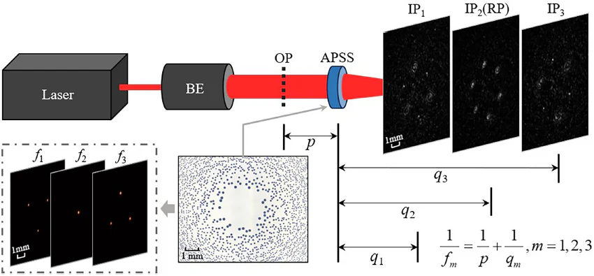
Figure1Schematic of the experimental optical path for single-exposure anisotropic array coherent diffraction imaging
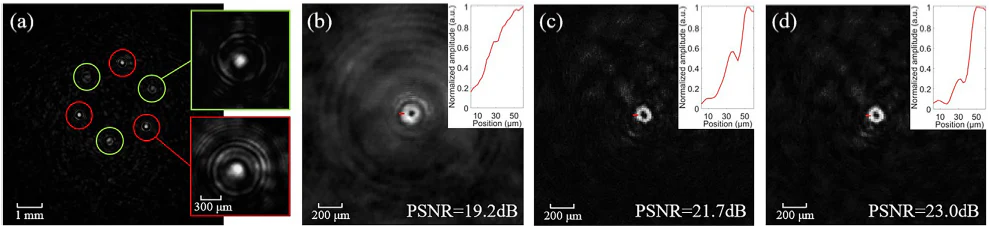
Figure2Experimental results(a)diffraction patterns, reconstructions under different combinations(b)one pair,(c)two pairs,(d)three pairs
Source: Shanghai Institute of Optics and Fine Mechanics (SIOM) · Research Updates · siom.cas.cn