How the Mantis Shrimp's Eye Detects Color
How the Mantis Shrimp Eye Senses Color
Topics and contents
Topic
On this page
The research team analyzed 17 visual pigments in the mantis shrimp eye and found that two red-absorbing rhodopsins can be switched on and off by red and blue light. In total, four rhodopsins showed bistability, and two additional blue-light rhodopsins could be switched by blue and orange light. The study was published in Sci. Adv. (doi: 10.1126/sciadv.aef5666). After optimization, these receptors could be used as optogenetic molecular switches to study human G signaling pathways.
Two red-light receptors can be switched between states by different wavelengths, suggesting that mantis shrimp visual proteins could serve as reversible molecular switches in studies of optogenetic signaling pathways.
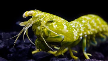
Researchers analyzed 17 visual pigments in the mantis shrimp’s complex eye, discovering two red-light-absorbing proteins. [Image: Michaël Bok]
Mantis shrimp are known to possess one of the most unusual and complex visual systems in the entire animal kingdom. Their highly mobile eyes have at least 12 types of photoreceptor cells, compared to just four in humans, and some species can even detect circularly polarized light.
Now, an international team of researchers has analyzed 17 visual pigments in the mantis shrimp eye involved in color sensing, discovering two red-light-absorbing proteins that can switch on and off using different wavelengths of light (Sci. Adv., doi: 10.1126/sciadv.aef5666). Efficient red-light detection is remarkably rare in animals, and these proteins could potentially serve as molecular switches to modulate disease signaling pathways in optogenetics experiments.
Far-red sensitivity
Visual pigments, housed within the photoreceptors of the eye, help animals detect light by transforming light energy into electrical signals. They consist of a light-sensitive chromophore and a protein called opsin, with the visual pigments in rod photoreceptor cells known as rhodopsins. The researchers aimed to uncover how multiple rhodopsins contribute to color sensing ability in the mantis shrimp species Neogonodactylus oerstedii, found in the Caribbean.
“The motivation for the study was curiosity—exploring the functionality and role of the highest number of rhodopsins found in an invertebrate animal eye thus far,” said study author Marjorie Liénard, a researcher at GIGA-ULiège, Belgium.
Liénard and her colleagues measured the light sensitivity and signal-transmission mechanisms of the rhodopsins in vitro, outside the tremendous complexity of the eye, selecting 17 to analyze mostly from the midband eye region. This region has six rows of ommatidia, or clusters of photoreceptor cells, with rows one to four mediating color discrimination and rows fine and six being responsible for polarization vision.
“In addition to numerous green rhodopsins similar in spectral sensitivity to our human green cone rhodopsin, three are absorbing at the red edge of the visible spectrum, and among them two are bistable—that is, they can switch on and off with red and blue photons of light,” Liénard said.
Downstream applications
A total of four rhodopsins exhibited bistability, demonstrating the capacity for reversible, light-driven transitions between two stable states without requiring an external cellular mechanism to regenerate. In addition to the two red rhodopsins, two blue rhodopsins could switch on and off with blue and orange light.
The bistability suggests that these light receptors function and reset rapidly, possibly to accommodate quick underwater fluctuations in light abundance and spectrum. Interestingly, the opsins of the mantis shrimp belong to the family of G-protein-coupled receptors (GPCRs), the largest family of human membrane proteins and an important class of drug targets.
“These light-sensitive receptors couple to a number of G-signaling proteins homologous to those found in many human tissues and cell types,” said Liénard. “With potential optimizations, they may present interesting features for the study of human G-signaling pathways. Red light penetrates tissues a little bit deeper than blue light, and this could be an advantage for specific downstream applications.”
Source: Optics & Photonics News · optica-opn.org