Xi'an Institute Advances Microscopic Imaging Through Scattering Media
中国科学院西安光机所在散射显微成像技术领域取得新进展
Topics and contents
The Ultrafast Optics Laboratory at the Xi'an Institute of Optics and Precision Mechanics has made new progress in optical microscopic imaging through scattering media.
The original article provides specific data and details that help gauge actual progress in this direction
Recently, the Transient Optics Research Laboratory at the Xi'an Institute of Optics and Precision Mechanics (XIOPM), Chinese Academy of Sciences, made new progress in optical microscopy through scattering media. The findings were published in Journal of Physics: Photonics. The co-first authors are Associate Researcher Peng Tong and Assistant Researcher Li Runze of XIOPM, and the corresponding authors are Researcher Dan Dan and Researcher Yao Baoli of XIOPM.
With its unique advantages of being non-invasive and damage-free, optical microscopy is a core tool for observing microscopic life processes in life science research. However, the refractive index inside biological tissue is highly non-uniform, causing severe scattering and wavefront distortion as light passes through — like viewing the microscopic world through multiple layers of frosted glass. This not only blurs the image but also greatly limits imaging depth. Existing scattering compensation techniques generally suffer from complex systems, heavy computation and reliance on labels. Conventional transmission microscopy struggles to correct wavefront distortion in both the illumination and detection paths simultaneously, while wavefront shaping can focus light through scattering media but is limited by the relatively large focal spot, restricting resolution.
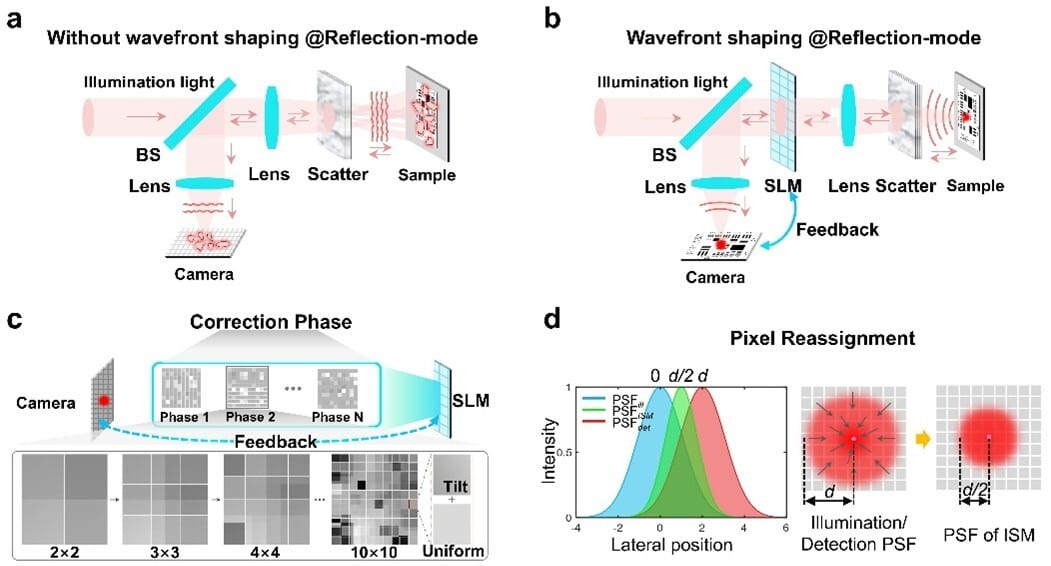
Figure 1: Principle of high-resolution imaging through multilayer scattering media via wavefront shaping and image scanning microscopy
To address this, the team proposed a guide-star-free, bidirectional wavefront correction imaging technique. Using light reflected from the sample as the feedback signal, a single spatial light modulator compensates wavefront distortion in both the incident illumination and the returning detection path, enabling non-invasive optical refocusing without fluorescent labels.
The technique combines wavefront shaping with image scanning microscopy, further reducing the focal spot size beyond scattering focusing. A correction strategy that progressively increases the number of control units, together with a layered scattering compensation mechanism, greatly improves computational efficiency while maintaining accuracy.
Experimental results show that the technique achieved high-quality focusing in a three-layer scattering medium with a total optical thickness of 1.82. Combined with pixel reassignment and deconvolution processing, the system resolution improved by about 1.76 times compared with scattering correction alone.
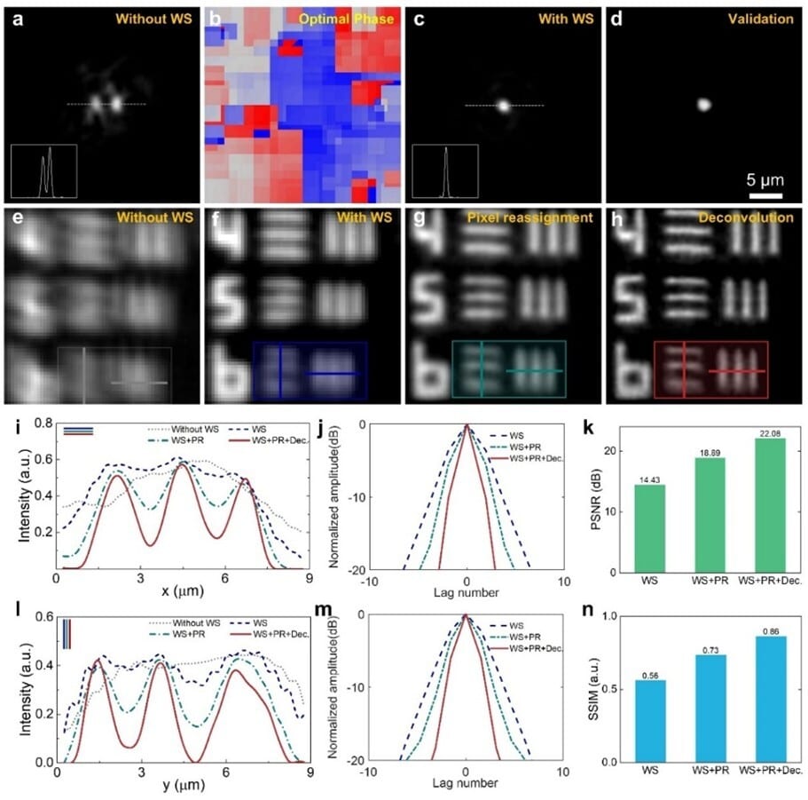
Figure 2 High-resolution imaging results through scattering media
The technique addresses the difficulty of simultaneously compensating for scattering distortions in both illumination and detection paths in optical microscopic imaging, opening a new route to deep-tissue, high-resolution imaging of biological samples. It is expected to find future applications in biomedical testing and related fields.
Peng Tong explained, "Scattering of light by biological tissue is the main obstacle to deep, clear imaging with optical microscopes. By precisely controlling the optical field, we restore order to the scattered light, effectively overcoming interference along the light propagation path, achieving a breakthrough from 'invisible' to 'visible' and then to 'clearly visible.'"
The research was supported by the National Natural Science Foundation of China, the National Key R&D Program of China, and the Shaanxi Provincial Key R&D Program, among other projects.
In recent years, the team of Researcher Yao Baoli at the Xi'an Institute of Optics and Precision Mechanics (XIOPM) has conducted in-depth research in imaging through scattering media and optical microscopic imaging, achieving a series of advances, including imaging through dynamic scattering media, non-invasive microscopic imaging through scattering media, and two-photon adaptive optics imaging. Related results have been published in journals such as Nanophotonics, Photonics Res., Appl. Phys. Lett., Opt. Express.
Source: Xi'an Institute of Optics and Fine Mechanics (XIOPM) · Research Progress · opt.cas.cn