XIOPM Makes Progress in Label-Free 3D Microscopic Imaging
Topics and contents
A team led by Yao Baoli at XIOPM has made new progress in label-free 3D microscopic imaging.
The source provides specific data and details that help assess actual progress in this direction
Recently, a team led by researcher Yao Baoli at the Ultrafast Optics Research Laboratory of the Xi'an Institute of Optics and Precision Mechanics (XIOPM), Chinese Academy of Sciences, made new progress in label-free 3D microscopic imaging. The findings were published in Journal of Physics: Photonics. The first author is Li Yan, a 2026 Ph.D. student at XIOPM; the corresponding authors are researchers Min Junwei and Yao Baoli. XIOPM is the first affiliation and corresponding institution.
Optical diffraction tomography (ODT) is a label-free, non-invasive, quantitative 3D microscopic imaging technique that can reconstruct the three-dimensional refractive index distribution inside transparent or semi-transparent samples. It has important applications in cell biology, histopathology, microbiology, and materials science. However, conventional ODT systems typically rely on a Mach-Zehnder interferometer configuration, highly coherent laser sources, and mechanical scanning devices, making them complex and demanding in terms of stability. Intensity-based ODT methods using LED arrays, which have emerged in recent years, simplify the hardware but generally require multiple intensity images and complex iterative algorithms to recover the phase, limiting imaging speed and reconstruction efficiency.
To address this, the team proposed an optical diffraction tomography method based on programmable LED array illumination and a quantitative phase camera (LED-ODT). The method modifies a standard commercial microscope, using a programmable LED array to achieve fast, non-mechanical multi-angle illumination. With the team's in-house-developed quantitative phase camera, the Q-camera, the complex amplitude of the sample under single-angle illumination can be obtained in a single exposure. The multi-angle complex amplitude data are then mapped into the 3D frequency domain using the Rytov approximation and the Fourier diffraction theorem, ultimately reconstructing the sample's 3D refractive index distribution (Figure 1).
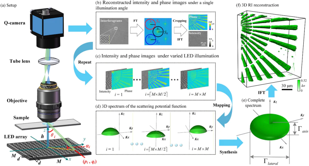
Figure 1. Schematic of the LED-ODT system and its working principle
Based on the LED-ODT method, the team built a compact label-free 3D microscopic imaging system consisting of a 15×15 programmable LED array and a Q-camera. The system achieves a maximum illumination angle of 52.9° and can acquire interferograms at 225 illumination angles within 2.7 seconds. The technique combines the complex amplitude measurement accuracy of conventional interferometric ODT, the phase measurement stability of common-path ODT, and the hardware simplicity and ease of operation of intensity-based ODT, delivering accuracy, stability, and usability in one package.
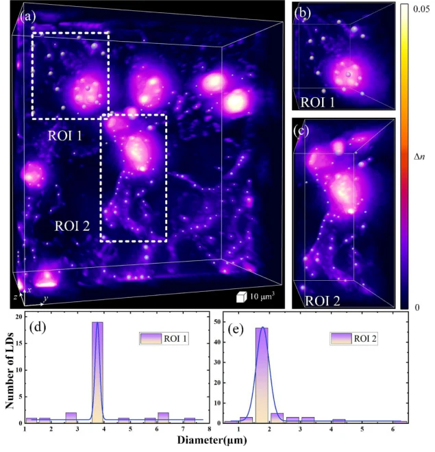
Figure 2. 3D refractive index reconstruction results for COS-7 cells
Experimental validation showed that the method delivers excellent 3D refractive index imaging of unstained COS-7 cells and water-soaked sterculia seeds. It clearly resolves subcellular structures such as lipid droplets within COS-7 cells (Figure 2), while also revealing the spatial heterogeneity of the 3D refractive index distribution inside water-soaked sterculia seeds (Figure 3), offering a new quantitative 3D imaging tool for studying the structure and function of natural biomaterials.
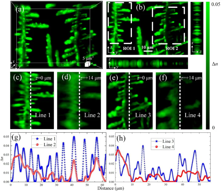
Figure 3 3D refractive index reconstruction of a water-soaked sterculia seed
Li Yan explains: "Put simply, this is like taking a contactless 3D full-body X-ray of a microscopic sample. It is as if 225 mini fill lights were arranged around the sample and switched on one by one from different angles. There is no need to move the machine or adjust the sample back and forth; in just 2.7 seconds, the fine internal structure is captured clearly. It can also be mounted directly on a standard microscope, so the barrier to use is very low."
LED-ODT is compact, low-cost and easy to operate, and can be integrated directly with standard optical microscope platforms. While maintaining imaging quality, it greatly improves the practicality of ODT technology, providing a fast, label-free, high-quality 3D quantitative microscopic imaging tool for observing transparent biological samples, analyzing cell metabolism, studying tissue pathology and characterizing natural polymer materials.
The work was supported by the National Natural Science Foundation of China, the National Key R&D Program of China, and the Youth Innovation Promotion Association of the Chinese Academy of Sciences.
Researcher Yao Baoli's team has long focused on label-free 3D microscopic imaging, accumulating extensive technical experience and achieving numerous research results. The team has established in-depth collaborations with research institutes and universities in China and abroad, and has developed various label-free 3D microscopic imaging devices for applications such as flow visualization and industrial inspection. The team has published more than 300 papers in journals including Optica, PNAS, PRL, Rep. Prog. Phys. and Adv. Opt. Photon., been granted multiple national invention patents, and received honors including first and second prizes for Science and Technology of Shaanxi Province and the Shaanxi Key Science and Technology Innovation Team award.
Source: Xi'an Institute of Optics and Fine Mechanics (XIOPM) · Research Progress · opt.cas.cn